
Digital X-ray Exam
Dental x-ray are the one of the diagnostic tool to see internal dental structure and surrounding structure like Dental caries, infection and bone condition etc.
It’s possible for tooth decay, apical root infection, defective restoration, periodontal disease, etc. to be missed during a clinical exam, and radiographic evaluation of the dental and periodontal tissues is a critical segment of the comprehensive oral examination.
Benefits of a Digital X-ray System:
- The images are displayed instantly.
- An enhanced, larger, clearer picture than conventional x-ray resulting in better diagnostic outcome.
- It can be recorded as a digital file which is easy and convenient for tracking.
- Better for the environment because there is no use of toxic processing chemicals or lead foil that are in the conventional dental film system.
- Produces a fraction of the radiation required for the conventional x-ray system.
Type of dental x-ray:
1) Extraoral x-ray:
1.1) OPG
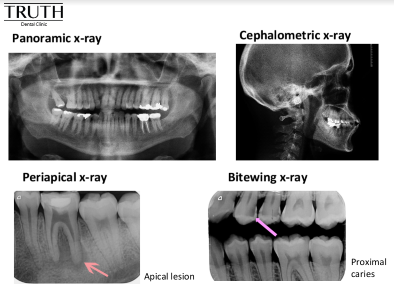
An OPG is a panoramic or wide view x-ray of the lower face. All the teeth of the upper and lower jaw are presented on a single film.
Benefit:
- A general overview of the teeth and the bone which supports the teeth.
- It demonstrates the number, position, and growth of all the teeth including those that have not yet surfaced or erupted.
- Jawbone problem detection.
- Temporomandibular joint, or TMJ, evaluation.
- Orthodontic treatment planning.
- Wisdom teeth assessment.
- Maxillary sinus evaluation.
- Mandibular nerve evaluation.
1.2) Lateral Cephalometric x-ray:
A Ceph or Lat Ceph is a lateral or side view x-ray of the face, which demonstrates the bones and facial contours in profile on a single film. This type of x-ray is commonly used in orthodontic treatment diagnosis and planning.
2) Intraoral x-ray:
2.1) Bitewing x-ray
Benefit:
- Interproximal caries
- Recurrent caries under existing restorations.
- Bone support determination in extensive bone loss case
2.2) Periapical x-ray:
Benefit:
- Show the whole tooth ; from the crown, to the root area where the tooth attaches into the jaw.
- Each film shows all teeth in one portion of either the upper or lower jaw.
- Use to detect any unusual changes in the root and surrounding bone structures.