
Root Canal Therapy
Our teeth are not only a single layer. In fact, they consist of three layers: “Enamel” First layers) “Dentine” (second layers ) and “Dental Pulp” (Third layers).
A dental pulp contains nerve and blood vessels. When the infection occurs at the pulp or third layer of a tooth, at that time tooth need root canal treatment or Endodontic treatment. Pulp infection is caused by bacteria that live in the mouth and invade the tooth.
Causes of dental pulp damage:
- Deep dental caries
- Traumatic force
- Broken tooth from accident
- Tooth displacement from accident
- Gum disease
- Chronic infection from adjacent teeth
- Tooth wear from chronic mechanical or chemical damage
- Intentional tooth preparation for cosmetic treatment
Treatment processes:
- Access opening
- Pulp or nerve removal or existing root canal filling removal
- Root length measurement by apex locator and x-ray machines
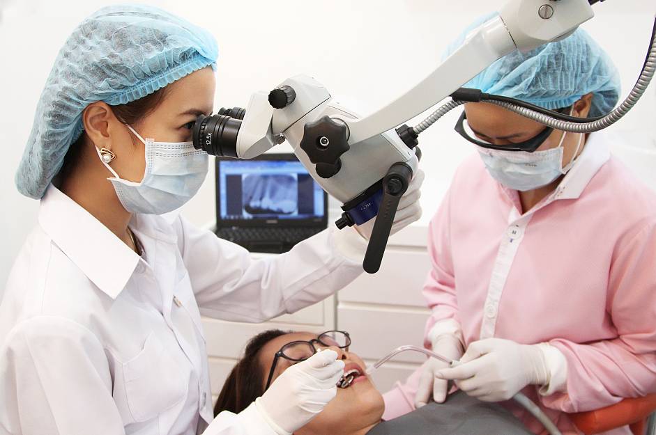
- Mechanical instrumentation by endodontic files and rotary instrument. This is a an important step which should be done by dental microscope. This will help prevent possible complication. The endodontic files will clean and smooth out root canal wall to eliminate all the bacteria accumulation.
- Root canal medicament consideration
- Root canal filling by system B obturation system and root canal filling material If endodontic treatment or first root canal treatment was done in the past and there remains root infection, endodontic retreatment or root canal retreatment is recommended. A typical cause of remaining root canal infection is from untreated root canal. Dental microscope will help to magnify the root canal Mostly, root canal retreatment requires a step of root canal medication for at least a week prior to the root canalobturation.
Number of treatment visits and duration:
| Condition | Number of visits |
| Non apical infection tooth | Single visit |
| Intentional root canal treatment | Single visit |
| Apical infection tooth | At least 2 visits with 1 week apart |
| Root canal retreatment | At least 2 visits with 1 week apart |
At Truth dental clinic, Root canal treatment will is performed under OPMI dental microscope by skillful root canal specialist or endodontist.
In the 21st century the OPMI plays a vital role in endodontics and endodontists have led the way in embracing the OPMI into daily clinical practice. Endodontic treatments can be very challenging due to the complexity of the anatomy of the root canal system. In the past root canal treatment was performed predominantly by feel. With the aid of the OPMI, structures can be seen that remain hidden to the naked eye and treat- ment can be carried out with far greater precision and predict- ably than ever before. Microscopy in endodontics has become a way of life
Benefit of having OPMI microscope in root canal treatment
- Examination of the external surface of the tooth
- Removal of coronal restorations
- Preservation of tooth structure
- Identification of the floor of the pulp chamber
- Location of sclerosed canals
- Identification and orientation of curvatures in the radicular access
- Identificatio of internal cracks, ledges and blockages in the root canal
- Evaluation of canal cleanliness after preparation
- Ensure optimal obturation
- Assessment of existing root fi lings
- Identificatio and management of perforations
- Assessment of coronal leakage
- Evaluation of endodontic instruments after use
- Removal of root fi ling materials in non-surgical re-treatment
- Management of soft and hard tissues in surgical re-treatment